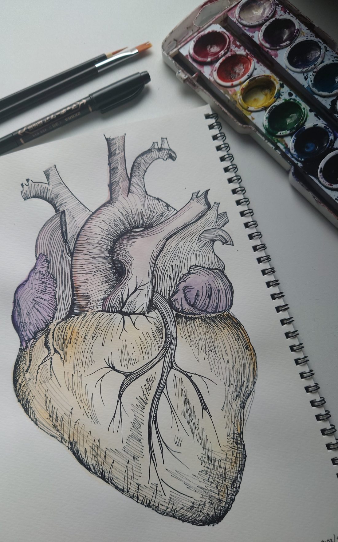
A tactful method of developing one’s understanding of the human heart’s anatomy is through drawing it! One study reported that “when studying anatomy science, drawing is one of effective important methods because it is an integration of ideas and knowledge of vision thereby increasing comprehension and learning motivation of college students” (Joewono et al. 2018).
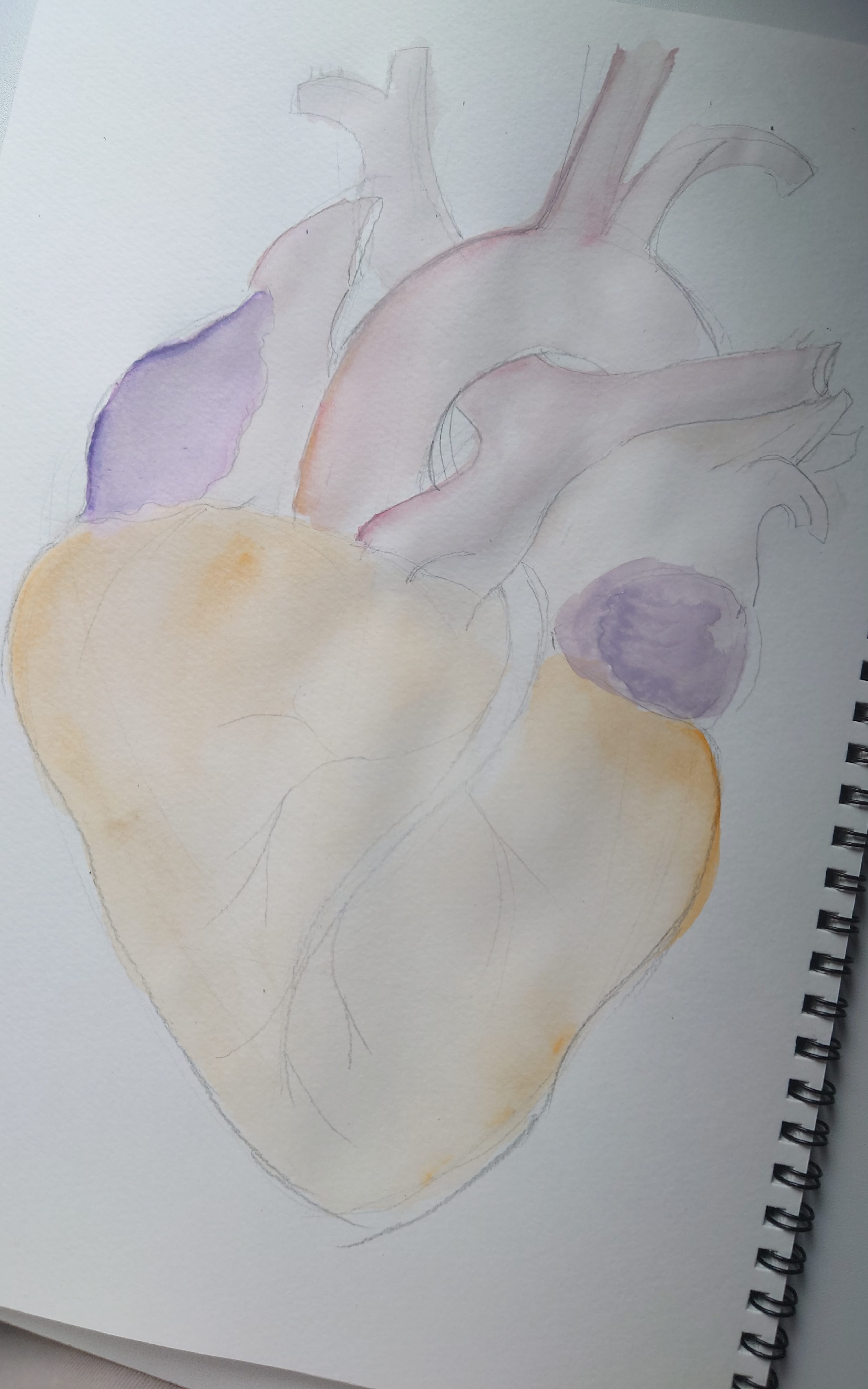
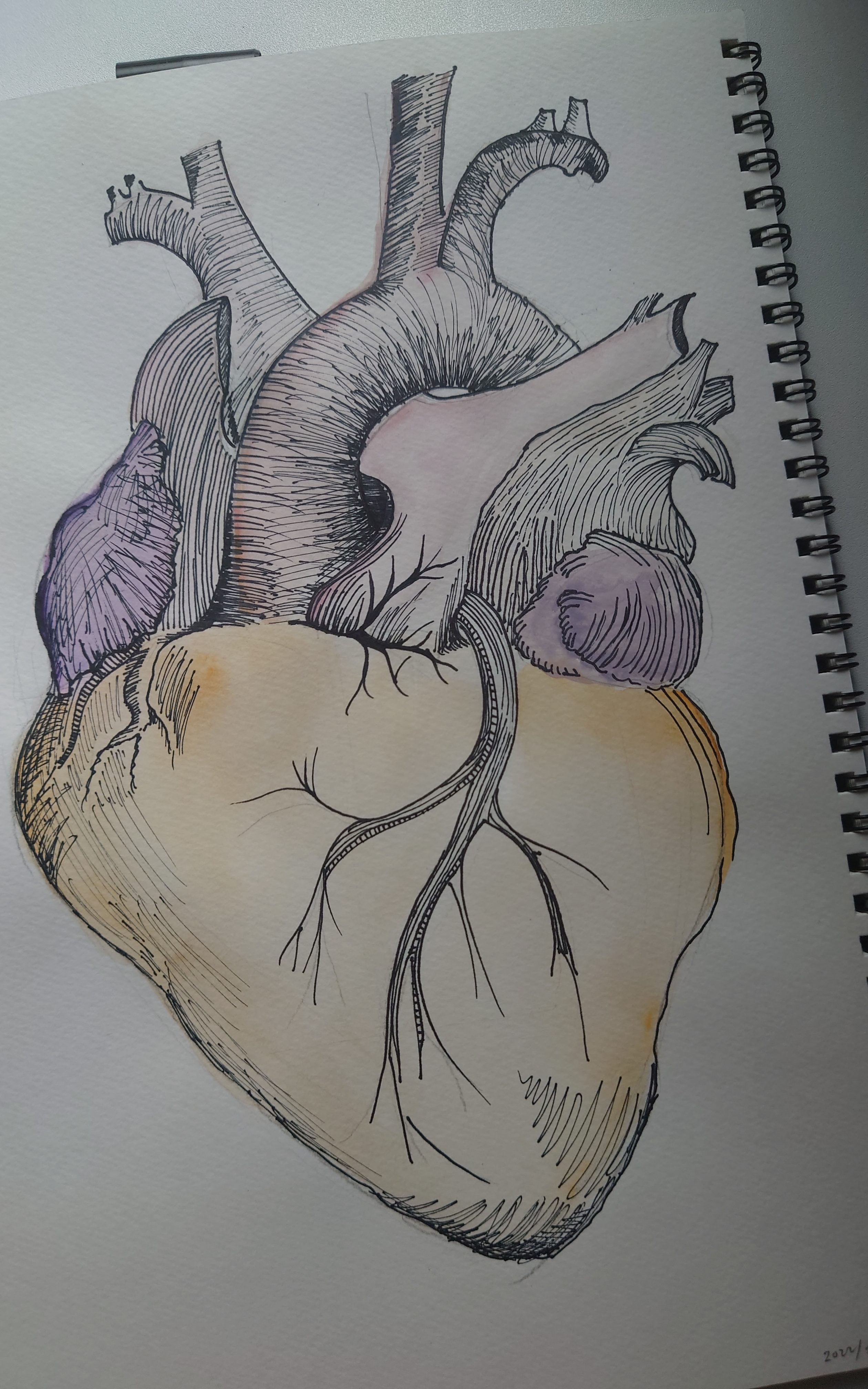
In my personal test of these findings, I spent time on February 15th, 2022 to complete this watercolour and marker ink mixed media artwork of the human heart’s anatomy. The process began with a simple search for an accurate reference image to proportionately sketch out the heart and its main components. Next, I chose watercolour paints that resembled the hues found in Renaissance period artworks to highlight those main components. Once the paint dried, I used a black marker to outline the main components of the heart followed by the detail.
The process look about 3 to 4 hours one lonely afternoon and I managed to create something meaningful. For those of you who are students in the field of clinical and health sciences, you may use post-it notes to label over the parts of the heart to test your knowledge!
lf you enjoyed this artwork then give this post a like or let me know in the comments below! If you have any inquiries about my artwork, please contact me here. To follow my art on Instagram, click here.


References
Joewono, M, Karmaya, NM, Wirata, G, Yuliana, I, Widianti, GA & Wardana, NG 2018, ‘Drawing method can improve musculoskeletal anatomy comprehension in medical faculty student‘, Anatomy & Cell Biology, vol. 51, no. 1, pp. 14 – 18.
