


Leonardo Da Vinci was a master of nothing and jack of all trades. It is said that he invested his earnings from selling his fine art paintings into purchasing cadavers, dissecting them and accurately illustrating human anatomy1. Similar to Mr. Da Vinci, I also share a fascination for the magic and mystery of the human anatomy and its physiology. In this article, I illustrate the different steps to showcasing the beauty of what is inside us.
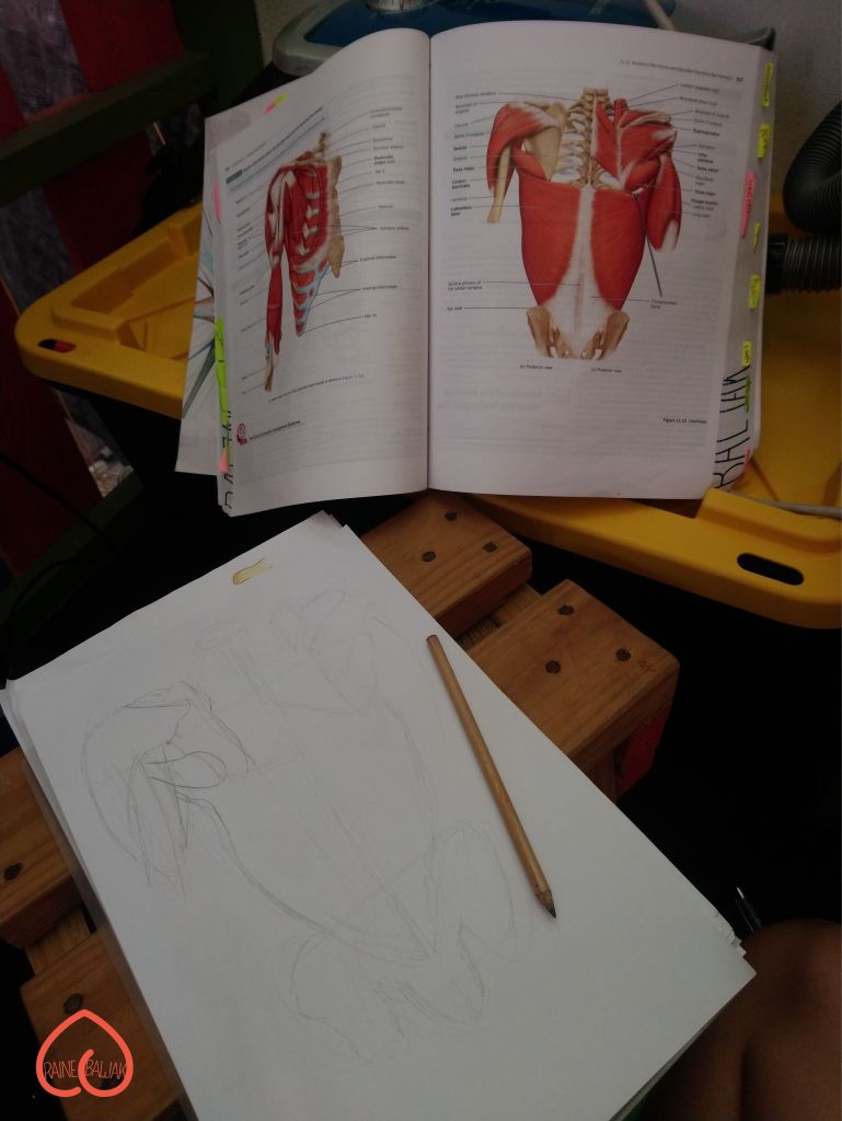


I drew these illustrations while studying human anatomy for a physical therapy degree at university. These illustrations were done to better comprehend each muscle, bone, ligament and joint that come together to form our bodies.


Knowing yourself is the beginning of all wisdom.
Aristotle


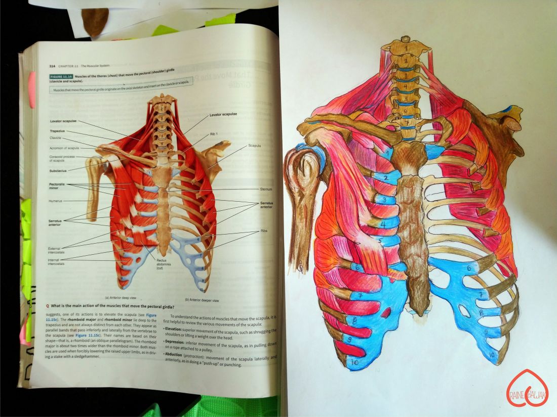
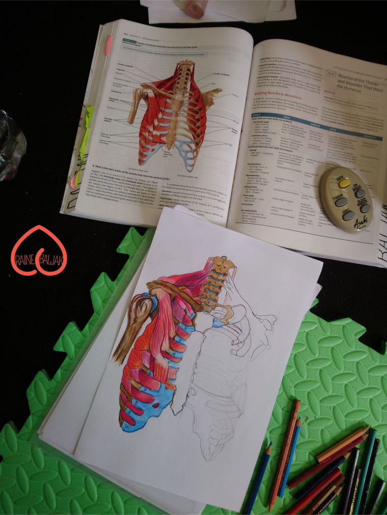
The above series of images were drawn with pencil, outlined in pen ink, then added vibrant hues in coloured pencils.
While these on the left, were illustrated strictly using coloured pencil to identify specific sections of muscle of the human body.

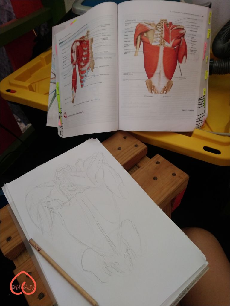
These last two illustrations, were done only using pen ink in the middle of a human anatomy lecture. The only visual references I had at the time were from images projected onto the board of the room.
The illustration on the left was made to identify facial bones, sutures and cranial sections. While the image on the right details the rib cage and vertebrae of the spine.
If you enjoyed my drawings, be sure to like this post. If you are interested in owning your own piece then follow this account.

Reference
- https://en.wikipedia.org/wiki/Leonardo_da_Vinci
- Gerard J. Tortora, Bryan H. and Derrickson 2017, Tortora’s Principles of Anatomy and Physiology, 15th Edition, Global Edition, United States of America.
